

3D X-Ray Microscope (MicroXCT-200)
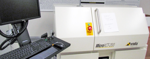
The National Institutes of Health (Shared Instrumentation Grant program) funded purchase of its first state-of-the-art high-resolution 3D X-ray Microscope, the MicroXCT-200, in Nebraska.
High spatial resolution pixel size down to 0.6 µm
The MicroXCT-200 system is available to meet the objectives of the scientific community interested in high resolution imaging of bone tissue, and materials that include muscle, other soft tissue, etc.
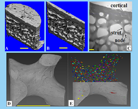
This MicroXCT-200 has been used recently to investigate osteocyte lacunar properties in bone tissue from humans (iliac bone biopsies representing estrogen loss, various treatments from drug trials), animal models representing various bone conditions (disuse, ovariectomy-related bone loss, PTH treatment, genetic regulation of bone mass, cochlear bone structure, embryonic skeletons, microdamage, etc.), and insects (studying detailed morphologies, etc.).
Use the MicroXCT200
We welcome use of the MicroXCT200 system by scientists from local institutions and from across the United States.
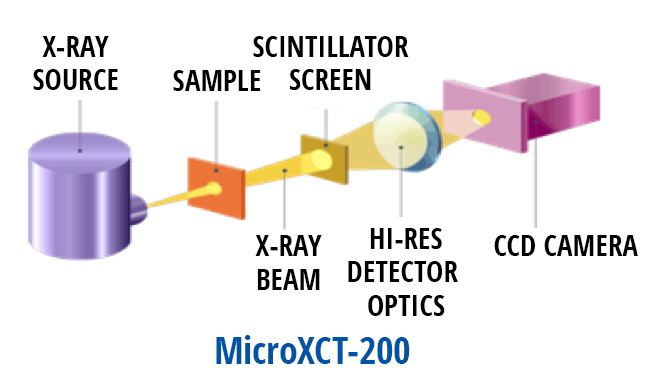
The MicroXCT-200 system is a 3D X-ray microscope developed by Zeiss/Xradia for industrial and academic research applications.
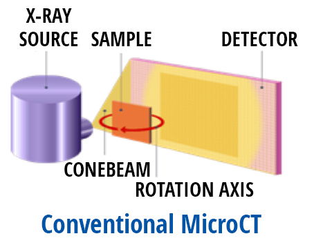
The MicroXCT-200 system provides excellent magnification, allowing a submicron level pixel resolution equivalent to examining 1/100th of the width of a single strand of hair. Such resolution has only been approached previously by synchrotron radiation (SR) microscopy.
However, unlike SR, the MicroXCT200 is a lab-based system readily available to a scientist without any wait time. The 3D X-ray microscope systems are also being used in the material science industry (geoscience, computer science, etc.)
Technical Information
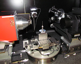
The MicroXCT-200 uses patented technology to acquire high quality images of specimens at an approximate highest pixel resolution of ~0.6µm, using tissue specimens measuring ~0.3 - 0.4mm in diameter.
Unlike existing lab based MicroCT systems, the instrument takes advantage of two stages of (geometric and light) magnification to produce enhanced 3D images providing sufficient details to characterize endpoints such as osteocyte lacunar characteristics; hair cells and the auditory epithelium of the inner ear; and the embryonic skeleton, using intact, non-artifacted samples of substantial size.
The Xradia systems convert the x-rays into light before they are recorded (note scintillator screen), then focus the light with a lens system that has various “objectives,” that permit studying small portions of whole specimens in 3D, just as if one was using a light microscope and rotating in a new objective. Then the image is recorded on a CCD camera, rather than a standard x-ray detector.
The core laboratories (Histomorphometry & Biomechanics) of Osteoporosis Research Center (ORC) follow principles of good laboratory practice (GLP), Quality assurance, data management and laboratory maintenance. The histomorphometry laboratory also maintains the CLIA certificate.
