Located in Room 209 of Criss I building, the facility is equipped with instruments commonly used for molecular biology and genomic experiments.
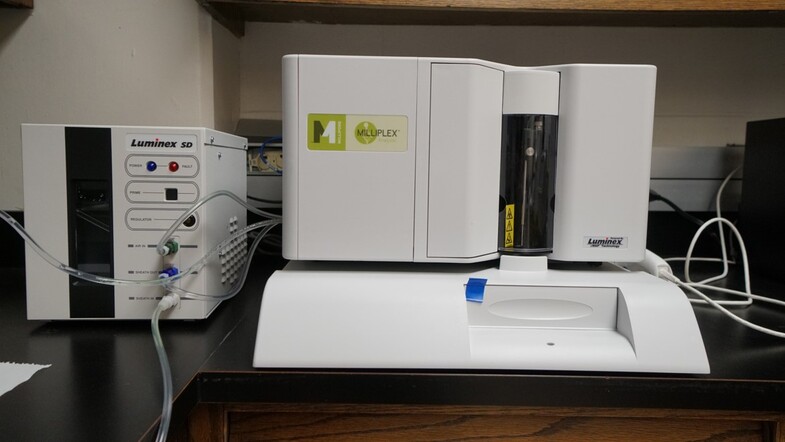
MILLIPLEX LUMINEX L200 ANALYZER
Milliplex Analyzer developed by Luminex uses multiplex xMAP bead technology that allows for teh simultaneous analysis of a broad selection of analytes (e.g. cytokines, hormones and adipokines) across a wide range of disease states, including metabolic disease, inflammation, neurodegenerative disease, toxicity, cancer and more.
Key Features:
Flow-Based Detection: Powered by xMAP® Technology, which uses a flow cytometry approach to identify and quantify individual microsphere sets, ensuring accurate multiplex results.
Versatile Multiplexing: Measures up to 100 targets in a single reaction, allowing users to conserve time, samples, and reagents. Compatibility with both magnetic (MagPlex®) and polystyrene microspheres offers flexibility for diverse assay needs.
High Throughput and Speed: Reads a 96-well plate in about 45 minutes (up to 12,800 tests per hour).
Broad Dynamic Range and Sensitivity: Greater than 3.5 logs dynamic range to detect low- to high-abundance targets. Sensitivity of fewer than 1,000 fluorochromes per microsphere supports the detection of subtle expression differences.
QUANTSTUDIO 3 REAL-TIME PCR
The Applied Biosystems™ QuantStudio™ 3 Real-Time PCR System is an accessible, high-performance benchtop instrument designed to empower researchers of all experience levels. Combining intuitive software, interactive touch-screen functionality, and secure connectivity via Thermo Fisher Connect, it enables quick setup, rapid data analysis, and easy collaboration.
Key Features:
Easy-to-use software and touch screen: An interactive interface and large, icon-driven touch screen guide you step-by-step through experiment setup and data analysis. Preoptimized protocol templates help eliminate guesswork, reducing time spent on training and troubleshooting.
Precise and reliable performance: With the ability to detect as little as 1.5-fold differences in target quantity in singleplex reactions and a broad dynamic range (up to 10 logs), the QuantStudio 3 system delivers the accuracy you need for gene expression, SNP genotyping, and other genomic applications.
Flexible experiment design: The system supports both probe-based (e.g., TaqMan®) and intercalating dye (e.g., SYBR™ Green) chemistries. Four optical channels allow for multiplexing of up to four targets in a single reaction, saving time and resources.
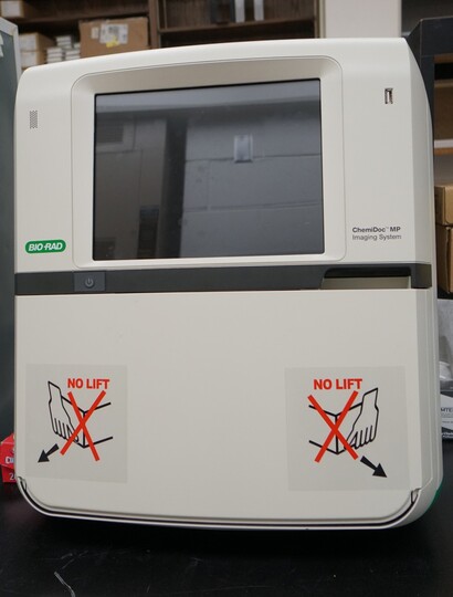
The ChemiDoc MP Imaging System is a full-feature instrument for imaging and analyzing gels and western blots. It is designed to address multiplex fluorescent western blotting, chemiluminescence detection, general gel documentation applications, and stain-free technology imaging needs. Features include:
- All-in-one flexible imaging - get precise, reproducible fluorescent, chemiluminescence, and colorimetric gel and blot detection, analysis and documentation in a single system
- Multiplex fluorescent western blotting - detect up to three proteins simultaneously and eliminate the need for stripping and reprobing
- Ease of use - features include automatic selection of optimal light source by application, auto focus, auto exposure, and preview features ensure optimal images. Learn in minutes with on screen help.
- Stain-free protein normalization - stain-free imaging permits the normalization of bands to total protein in both in gels and blots; eliminates the need for housekeeping proteins
- Sample Trays and Smart Tray Technology - three sample trays available to cover diverse imaging applications. Smart Tray Technology automatically recognizes the application-specific trays and adjusts imaging parameters and software options accordingly
QUANTSTUDIO ABSOLUTEQ DIGITAL PCR
Elevate your research with the QuantStudio™ Absolute Q™ Digital PCR System—a powerfully simple platform that integrates all steps of digital PCR into a single instrument. Proprietary microfluidic array plate (MAP) technology ensures over 95% of your sample is accurately measured, delivering precise, sensitive, and absolute quantification without the need for standard curves. From genomics, oncology to infectious disease, this versatile system is designed to accelerate your discoveries with minimal hands-on time and streamlined data analysis.
Key Features:
Single-Instrument Workflow: Combine reaction digitization, thermal cycling, and data acquisition in one compact system.
Microfluidic Array Plate Technology: Achieve consistent sample partitioning, with less than 5% dead volume and up to 20,000 micro-chambers per reaction.
High Sample Utilization: Analyze over 95% of your sample to boost sensitivity and accuracy, ensuring maximum data recovery.
Four-Color Multiplexing: Detect up to four targets in a single run, saving time and reagents while expanding your application range.
Broad Application Range: Ideal for oncology, reproductive health, infectious disease, inherited disease, gene expression, copy number variation, and more.
User-Friendly Software: Easily set up plate layouts and thermal conditions, then visualize results in clear, exportable formats for downstream analysis.
JESS CAPILLARY WESTERN
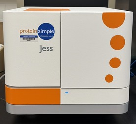
Meet Jess—the fully automated, next-generation protein analysis platform that brings together Western blotting into a single, easy-to-use system. Designed to eliminate tedious steps and human error, Jess streamlines protein separation, immunodetection, and data analysis into an efficient, three-hour workflow. Simply load your samples and reagents, and let her handle the rest. Her high-sensitivity chemiluminescent and NIR/IR fluorescence modules capture picogram-level signals, giving you reliable, publication-ready results without the guesswork.
Whether you’re monitoring protein expression, running quantitative immunoassays, or comparing multiple targets, Jess maximizes your data output with minimal sample input—perfect for precious or low-abundance samples.
Key Features:
Full Automation: Jess handles protein separation, immunodetection, and data analysis—all in a single run—minimizing hands-on time and reducing user-dependent variability.
High Sensitivity: Achieve picogram-level detection with chemiluminescence or capitalize on superior fluorescence performance with NIR/IR modules for low-abundance targets.
Efficient Workflows: Run up to 25 samples per cartridge, and analyze results in as little as three hours.
Multiplex and Normalization: Detect multiple proteins simultaneously and use built-in total protein or fluorescent normalization to ensure consistent, reproducible results.
Streamlined Data Analysis: Easy-to-use software delivers both qualitative (Western blot–like) and quantitative (ELISA-like) insights, simplifying protein sizing, concentration measurements, and comparative analyses.
RePlex Capability: Quickly swap between different immunoprobes or total protein detection in a single run, maximizing data acquisition from limited or precious samples.
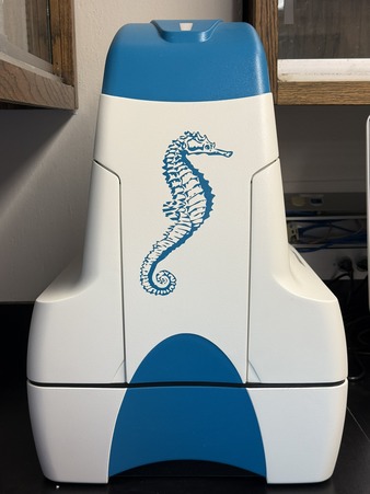
AGILENT SEAHORSE XFe24 FLUX ANALYZER
Experience a deeper understanding of cellular metabolism with the Agilent Seahorse XFe24 Analyzer—the trusted platform for real-time measurement of mitochondrial respiration and glycolysis in live cells. This high-sensitivity, 24-well system enables precise metabolic assessment of diverse samples, including islets, organoids and small organisms such as zebrafish larvae, C. elegans that require a larger well format. By simultaneously monitoring oxygen consumption and extracellular acidification, the Seahorse XFe24 delivers critical insights into how cells generate and expend energy.
The Seahorse XFe24 Analyzer simplifies everything from routine assays to advanced metabolic investigations, helping researchers uncover the mechanisms behind disease, immunity, cancer, and more. Whether you’re optimizing new treatments or exploring fundamental cellular processes, the Seahorse XFe24 puts the power of real-time metabolic data at your fingertips—empowering you to make breakthrough discoveries faster and with greater confidence.
Key Features:
24-Well Format: Perfect for working with specialized cell types, including islets and small organisms, that require a larger well size than traditional microplate formats.
Real-Time Metabolic Analysis: Monitors both oxygen consumption and extracellular acidification simultaneously, providing a complete picture of mitochondrial respiration and glycolysis in live cells.
Automated Compound Injections: Allows up to four sequential reagent additions per well, enabling multi-step protocols and reducing manual handling for greater reproducibility.
High Sensitivity: Detects metabolic activity in as few as 5,000 cells per well, making it suitable for rare or precious cell samples.
Flexible Experimental Design: Compatible with both adherent and suspension cells, as well as isolated mitochondria, for diverse applications across research areas like cancer, immunology, and metabolic disorders.
Robust Data Analysis Software: Delivers intuitive, automated calculations of key parameters—such as basal respiration, ATP-linked respiration, and spare respiratory capacity—streamlining the path from raw data to meaningful insights.
*Directed by Sarath Vijayakumar


