

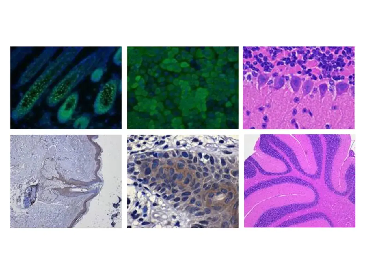
Histology Core
The Creighton University Histology Core Facility is a university-wide facility providing equipment and services to the research community. The CU Histology Core Facility (800 sf) provides services for the research community for studies utilizing animal and/or human tissues:
- Sample Processing
- Embedding
- Sectioning
- Histochemistry
- Immunohistochemistry
- Imaging
Current Equipment in the Histology Core:
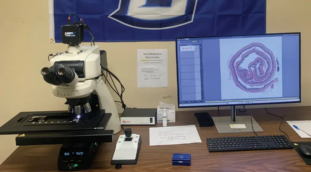
- Nikon BioPipeline Slide Scanner with Eclipse Ni-E Upright Microscope
- Olympus VS120 Virtual Slide Scanner ($10.00 per hour)
- Olympus Analysis Station (desktop computer)
- Nikon Eclipse Ci microscope with fluorescence & bright field applications
- Leica RM 2135 rotary microtome
- Finesse 325 rotary microtome
- Leica CM 1950 Cryostat
- Excelsior ES Tissue Processor
- Histostar Wax Embedding Center
- Gemini AS Auto Stainer for H&E staining
Nikon BioPipeline Slide Scanner
Features:
- Marzhauser 8-Slide Scanning Stage
- Basler Label Camera
- Basler BF Scan Camera
- Photometrics Prime BSI Express EPI Scan Camera
- NIS-Elements Slide Scanning Module
Nikon Eclipse Ni-E Upright Microscope
Features:
- Standard Ni-E hardware for Fluorescence and Brightfield illumination of cover-slipped slides (25 mmX75 mm, 1 in x 3 in)
- Nikon CF160 Plan Apo Lambda D objectives (2x, 4x, 10x, 20x, 40x)
- NIS-Elements Software and workstation
Image
Histology Services
Services Provided Include:
- Tissue processing
- Embedding
- sectioning
- Hematoxylin and eosin staining
- Immunohistochemistry
- Immunofluorescence
- Microscopy
The facility also provides equipment for researchers to use on a first come, first served basis. This equipment includes microtomes for paraffin sections, a cryostat for frozen sections, a vibratome for plastic sections, and a Nikon Eclipse Ci-L microscope for brightfield and fluorescence applications.
The Histology Core is co-located with the Molecular Core in Criss III, room 382.
