Core Facilities
Research Core Facilities
Learn more about our facilities below.
Department of Biomedical Sciences
Advanced Microscopy Core
Contact: Dr. Michael Nichols (mnichols@creighton.edu)
The Advanced Microscopy Core facility is composed of multiple advanced research microscopes and image analysis tools. New users must receive official training and sign a user agreement prior to being granted access to the facility’s tools or its reservation system. We are happy to provide training as well as to consult with users about their experimental and analysis needs. Please inquire if you have specific questions about any of the equipment or services available through our facility. If you are interested in becoming a new user of the Advanced Microscopy Core facility, please contact the core manager, Anthony Stender, about training and access to the facility.
For detailed information regarding the Advanced Microscopy Core’s equipment and services, please check out our main webpage under the “Advanced Imaging Services” tab at: https://www.creighton.edu/medicine/departments/biomedical-sciences/translational-hearing-center/technology
Department of Biomedical Sciences and Translational Hearing Center
Auditory and Vestibular Electrophysiology Core
Location: Criss I Building, Room 304
Contact: Dr. Sarath Vijayakumar (SarathVijayakumar@creighton.edu)
Dr. Michael Nichols (mnichols@creighton.edu)
The Auditory-Vestibular Electrophysiology Core provides infrastructure and training to support laboratories associated with the Translational Hearing Center at Creighton University to conduct multidisciplinary auditory and vestibular research.
Services and Equipment
1. Non-Invasive Hearing Screening Tests
- Auditory Brainstem Response (ABR)
- Otoacoustic Emissions (OAEs)
2. Cochlear Potential Measurements
- Cochlear Microphonics (CM)
- Compound Action Potentials (CAP)
- Endocochlear Potentials (EP)

3. Zebrafish Inner ear and Lateral Line Microphonic Assessment
4. Vestibular Electrophysiology
- Vestibular Evoked Potentials (VEP)
- Vestibular Ocular Reflex (VOR)
5. Cortical Auditory Evoked Potentials (CAEP)
Department of Biomedical Sciences
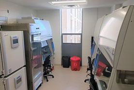
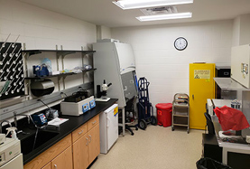
Cell & Tissue Culture Core
Location: Criss I Building, Room 402
Contact: Dr. Sarath Vijayakumar (SarathVijayakumar@creighton.edu)
Equipment
1. Cell Culture Core Equipment
- Biosafety cabinet: Axiom, Labconco C1 (x3)
- Incubator: PHCbi MCO-170AICUVL-PA cellIQ™ Series CO2 Incubator (x4)
- Benchtop Centrifuge: SORVALL ST8
- Microscope: Leica S40 microscope
- Automatic cell counter: Countess II (Invitrogen)
- Refrigerator
2. Tissue Culture Core Equipment
- Biosafety cabinet: Axiom, Labconco C1 (x2)
- Microscope: Leica Stereozoom S9D microscope
- Benchtop Centrifuge: SORVALL ST8, Eppendorf 5415D
- Incubator:
- Heracell Vios 160i
- Thermo Steri-cycle
- Refrigerator
Department of Biomedical Sciences
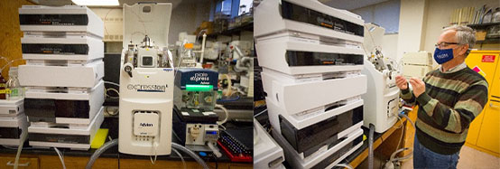
Mass Spectrometry Core
Location: Criss II Building, Room 303 & Beirne Research Tower, Lab 308
Contact: Dr. Molly McDevitt mollymcdevitt@creighton.edu
Equipment
1. Q Exactive Hybrid Quadrupole-Orbitrap Mass Spectrometer - Criss II, 303:
For the analysis of ligands, protein modifications and interactions, as well as small molecule and metabolomics studies. Sample delivery can be achieved using one of three methods:
- Direct Inject
- Vanquish UHPLC
- Easy-NanoLC 1200
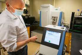
2. MALDI imaging: UltrafleXtreme MALDI-MS System - Criss II, 303:
- The innovative smartbeam-II™ laser enables ultra-high data acquisition speed in both MS and MS/MS at full systems performance. The well-established proprietary smartbeam laser provides unprecedented analytical and matrix flexibility in workflows from protein tissue imaging, intact proteins analysis, Glycoproteomics, biologics or oligo QC, to LC-MALDI proteomics - all fully enabled at 1-2000 Hz repetition rates.
- Bruker’s patented smartbeam technology is already widely accepted as the most viable MALDI imaging laser technology. The new ultrafleXtreme now enables low pixel sizes for high spatial resolution imaging. Importantly, outstanding spectral quality and signal intensity are maintained at even the smallest laser beam diameters.
- Broadband mass resolving power up to 40,000 enables precision proteomics via Bruker’s unique PAN™ technology for highest mass resolution across a very broad mass range, not just at a selected optimum.
- The novel FlashDetector™ combined with a minimum of 5 GS/s 10 bit digitizer and latest advances in electronics provide unmatched mass resolving power up to 40,000 and 1 ppm mass accuracy for highest confidence.
- The novel and unique laser-irradiation self-cleaning Ion Source ensures robust, long-term highest-performance operation. Very long MALDI laser lifetime in combination with automated source cleaning in just minutes leads to high uptime and low maintenance costs.
- Latest TOF/TOF technology: The high efficiency and sensitivity of the LID-LIFT process delivers MS/MS spectra with nominal mass resolution for peptides. Typically, full MS/MS data sets can be acquired with up to 1000 Hz laser repetition rate from low fmol levels within seconds. The Bruker bioinformatics software is perfectly adapted to analyze and visualize the match between the raw spectra and annotated peptide and protein structures.
- As a result, proteins can be identified with high confidence with unmatched ease.

3. Advion expressionL Compact Mass Spectrometer (CMS) - Beirne Research Tower, Lab 308:
- A single quadrupole mass spectrometer designed for the synthetic organic chemist. The instrument has unit resolution optimized for mass determinations of small molecules, peptides and proteins. Samples are introduced by either direct inject, after fractionation on an attached liquid chromatograph or by extraction from a developed thin layer chromatography plate using the Plate Express™ attachment.
Contact Us
Biomedical Sciences – MS & PhD Programs
Garrett Soukup
PhD Professor of Biomedical Sciences
garrettsoukup@creighton.edu
402.280.5754
Senior Program Coordinator
Sabina Coffiel
SabinaCoffiel@creighton.edu
402.280.3356